|
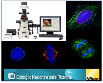
The laboratories of CNR-IBPM(Institute of Molecular Biology and Pathology ) @ San Lorenzo host the Nikon Reference Center for Central-Southern Italy, with last-generation widefieldlight microscopy equipment and advanced imaging software for automated image analyses. Imaging capabilities include:
-
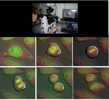
Time-lapse recording of live cells over several days,
-
Detecting intracellular molecular interactions,
-
Reconstructing cellular and subcellular structures at 3D level,
-
Obtaining quantitative information on the molecules or cells that are being imaged.
The Center is open to external users.
|
OUR EQUIPMENT
|
Up-right microcospe delivering crisp, clean and
true-to-life images of fixed fluorescent cell sample. |
Inverted microscope for
time-lapse imaging of living cells. |
|
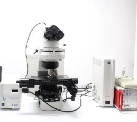
Nikon Eclipse 90i microscope
|
Nikon Eclipse Ti microscope
|
| Workstation |
| Nis-Elements HC 4.20 software (Nikon) for qualitative (deconvolution, 3D-projection, Extended Depth of Focus, LUTs, etc) and quantitative (measures of geometric features, intensity signals; object counts and tracking) image processing. https://www.microscope.healthcare.nikon.com/it_EU/products/software/nis-elements |
|
More on equipment...
|
| TIME-LAPSE IMAGING DATABASE |
|
Collection of individual entries by users, supporting both automatic and manual annotations, for standardization, comparison and retrieval of images, experiments and applications
|
| FIELDS OF APPLICATION |
|
The IBPM-Nikon microscopy platform supports versatile applications for both fixed and live cell samples and provides opportunities for coupling dynamic studies (time-lapse recording) with high resolution qualitative and quantitative image analysis of cells and cellular structures.
Applications include:
-
Real-time visualization of dynamic cellular processes (signaling, intracellular transport, organization of organelles and subcellular structures)
-
Single-cell analysis, to visualise cell heterogenity within a population
-
Recording of cellular morphological changes or cell death (in response to particular stimuli, damage, stress conditions)
-
Measurements of cell migration
-
High definition analysis of subcellular structures (5 fluorescence excitation channels, simultaneous visualization of 4 stainings, deconvolution)
-
Proximity ligation assays for in situ protein interactions and in situ post-translational modifications.
These applications are employed in the following fields:
-
Cell division and checkpoints
-
Inhibition of tumor cell growth
-
Differentiation of stem and progenitor cells
-
Validation of innovative drugs
-
The cell response to parasites, bacteria and viral infectious agents
-
Novel biocompatible matrices to support cell growth in studies of tissue regeneration
-
The interaction of nanoparticles with cells in toxicity and therapeutic studies
We are grateful to Assicurazioni Generali Spa, Fondazione Roma Terzo Settore (link is external) and Fondazione Monte dei Paschi di Siena (link is external) for their financial support in the set-up of the microscopy lab and to Regione Lazio - progetti LazioInnova for support to the implementation of facilities.
|
|